
The majority of patients who walk into a hair loss clinic in India have already decided, at least partially, what treatment they want. They have read about PRP, researched hair transplants, or been advised by a friend to try a particular supplement. They arrive with a treatment preference looking for confirmation, not with a clinical question looking for an answer.
The problem with this approach is that it skips the single most important step in any hair loss evaluation: understanding what is actually happening at the follicular level before recommending anything.
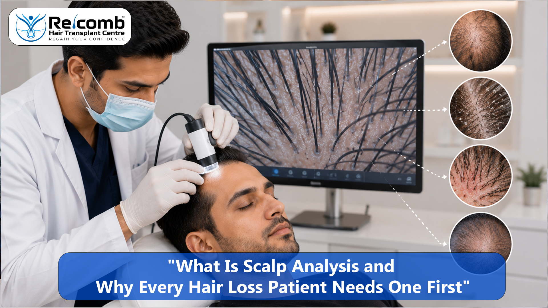
Scalp analysis is that step. It is the clinical examination of the scalp and its follicles using magnification, imaging, and structured assessment that gives the treating doctor specific, measurable information about what is driving the hair loss, how advanced it is, and which treatment has any biological basis for helping.
Without this information, every treatment recommendation is an educated guess. With it, the recommendation becomes a clinical decision.
If you have been losing hair and have not had a proper scalp analysis, a consultation at RECOMB is where that assessment starts.
Book a Scalp Analysis Consultation at RECOMB, Surat →
WhatsApp: +91 7624008000 | www.recombhair.com
What Scalp Analysis Actually Is
Scalp analysis is not a single test. It is a structured examination process that combines several components to build a complete clinical picture of the scalp and follicle environment. The core component is trichoscopy, a magnified examination of the scalp using a dermatoscope, but a complete scalp analysis extends beyond this to include a clinical history, scalp condition assessment, and in most cases targeted blood investigations.
Trichoscopy allows the clinician to examine the scalp at magnifications of 10 to 70 times or more, revealing structures and patterns invisible to the naked eye. At this level of magnification, individual follicle openings, hair shaft diameter, the presence of miniaturisation, scalp surface abnormalities, vascular patterns, and the early signs of scarring or inflammatory conditions become visible and measurable.
What the clinician sees through trichoscopy directly determines the diagnosis. A scalp that looks uniformly thin to the naked eye may show entirely different patterns under magnification depending on whether the cause is androgenetic alopecia, telogen effluvium, nutritional deficiency, scalp inflammation, or a scarring condition. Each produces a distinct trichoscopic signature that guides the clinical decision.
What Scalp Analysis Reveals That Naked Eye Examination Cannot
Follicle Miniaturisation
The defining feature of androgenetic alopecia at the follicular level is miniaturisation, the progressive reduction in hair shaft diameter over successive growth cycles driven by DHT. Under trichoscopy, this appears as a mixture of hair shaft diameters within the same scalp zone, with miniaturised hairs producing shafts visibly narrower than the normal-diameter hairs alongside them.
This variation in shaft diameter is the single most reliable trichoscopic sign of androgenetic alopecia. It is not visible to the naked eye or detectable by feel. It can be present and clinically significant in a patient whose scalp still looks reasonably full to external observation, which is why patients often present at a more advanced stage of miniaturisation than they realise.
Identifying miniaturisation through trichoscopy also quantifies its extent: what proportion of follicles in a given zone are miniaturised, how severely, and whether the miniaturisation is predominantly affecting the frontal zone, the crown, or both. This information directly determines the urgency of medical management and the viability of surgical planning in each zone.
Donor Density Measurement
For any patient considering hair transplant surgery, donor density measured through trichoscopy is the foundation of the entire graft plan. The number of follicular units per square centimetre in the safe donor zone, the average number of hairs per follicular unit, and whether the donor zone itself shows any early miniaturisation all determine the lifetime graft budget available to the patient.
This measurement cannot be accurately estimated visually. A donor zone that appears dense may have lower follicular unit density than expected under trichoscopy. One that appears average may have superior density that allows a more ambitious surgical plan. Without this measurement, graft recommendations are commercial estimates rather than clinical calculations.
Scalp Condition and Inflammation
Trichoscopy reveals the condition of the scalp surface and the perifollicular tissue in ways that standard examination cannot. Active seborrheic dermatitis appears as yellowish scaling around follicle openings with perifollicular redness. Early scarring alopecias such as lichen planopilaris show characteristic perifollicular whitening and scaling that is a specific and important early sign. Follicular plugging from hard water deposits or product buildup is visible as casting around follicle openings.
Each of these findings has specific treatment implications. A patient with active seborrheic dermatitis identified through trichoscopy before surgery is directed toward antifungal and anti-inflammatory treatment before any procedure is planned, because operating on an inflamed scalp compromises graft survival. A patient with early lichen planopilaris is directed toward urgent dermatological management to halt the scarring process before irreversible follicle destruction has occurred. Neither of these diagnoses would have been made reliably from visual examination alone.
Empty Follicle Openings and Scarring
Trichoscopy can identify follicle openings that are present but empty, meaning the follicle structure remains but is no longer producing a hair shaft. This distinguishes follicles in the telogen phase from those that have been permanently damaged. In a patient with suspected scarring alopecia, the pattern of empty follicle openings and the presence or absence of the follicle opening itself tells the clinician whether damage is ongoing, how extensive it is, and whether any follicles remain salvageable.
Hair Cycle Assessment
The proportion of follicles visibly in the telogen phase at the time of examination provides information about whether active, ongoing shedding is occurring beyond the baseline normal range. An elevated proportion of telogen hairs across the scalp, in the absence of the miniaturisation pattern of androgenetic alopecia, points toward telogen effluvium as the primary or significant contributing diagnosis.
Why Skipping Scalp Analysis Costs Patients More Than They Realise
The consequences of proceeding without a scalp analysis fall into two categories: wrong treatment and wrong timing.
Wrong treatment is the most direct cost. A patient diagnosed with hair loss on the basis of visual examination alone who receives PRP therapy may have androgenetic alopecia at a stage where PRP can contribute meaningfully, or they may have telogen effluvium where addressing the trigger would produce full recovery without any treatment, or they may have early scarring alopecia where PRP is inappropriate and urgent dermatological management is required. The visual presentation alone cannot distinguish these. The money spent on the wrong treatment is rarely recoverable.
Wrong timing is the less obvious but potentially more significant cost. A patient who undergoes a hair transplant without trichoscopy-based donor density measurement receives a graft plan built on estimates. Those estimates tend to over-recommend graft counts for commercial reasons, depleting the donor area at a rate faster than necessary for the patient's actual coverage needs. A patient who starts medical management for what is assumed to be androgenetic alopecia when the actual driver is a nutritional deficiency waits months for a result from treatment that cannot address the cause.
In every one of these scenarios, a scalp analysis performed before any treatment decision would have produced a different and better outcome.
What a Complete Scalp Analysis at RECOMB Involves
At RECOMB, scalp analysis is the first step of every new patient assessment before any treatment is discussed. The components are consistent across all patients.
Trichoscopy of the recipient zones identifies miniaturisation pattern, shaft diameter variation, follicle density, scalp surface condition, and the presence of any inflammatory or scarring signs. Trichoscopy of the donor zone measures follicular unit density, average hair count per unit, and donor zone health.
Norwood or Ludwig staging is performed using the trichoscopy findings alongside visual examination to formally document the current extent of hair loss.
Scalp condition assessment notes any seborrheic scaling, follicular plugging, perifollicular inflammation, or barrier disruption that requires treatment before or alongside any other intervention.
Blood investigation recommendations are made based on the trichoscopic findings and clinical history, covering at minimum serum ferritin, thyroid function, vitamin D, and complete blood count, with additional panels where the clinical picture suggests hormonal or autoimmune contributors.
The findings from all of these components are discussed with the patient in detail before any treatment recommendation is made, so the patient understands specifically what is driving their hair loss, how advanced the relevant processes are in each scalp zone, and what the clinical rationale is for whatever treatment is proposed.
Understand What Is Actually Happening on Your Scalp at RECOMB →
WhatsApp: +91 7624008000 | www.recombhair.com
RECOMB's Approach (2026)
At RECOMB Hair Transplant Centre, Surat, no treatment recommendation, surgical or non-surgical, follows a consultation that has not included a proper scalp analysis. Dr. Krishna Bhalala and Dr. Nilesh Kachhadiya treat scalp analysis as the non-negotiable foundation of every patient interaction, because a recommendation made without it is not a clinical recommendation. It is a commercial guess.
Patients who have been recommended treatments elsewhere without a scalp analysis are assessed from the beginning at RECOMB, regardless of what they were told previously. The findings from the RECOMB assessment either confirm the previous recommendation with specific clinical evidence, which increases the patient's confidence in proceeding, or identify discrepancies that change the treatment decision before any irreversible step has been taken.
Final Takeaway
Scalp analysis is not a premium add-on for patients who want extra information. It is the clinical minimum for any honest hair loss evaluation. Without it, the diagnosis is incomplete, the treatment recommendation lacks a specific foundation, and the patient is making a financial and medical commitment based on incomplete information.
Every patient experiencing hair loss deserves to know specifically what is happening at the follicular level before spending money on any treatment. That knowledge comes from a scalp analysis, not from a visual examination or a questionnaire.
Dr. Krishna Bhalala and Dr. Nilesh Kachhadiya conduct a limited number of personal consultations each week at RECOMB, Surat. If you have not had a proper scalp analysis and want to understand what is actually driving your hair loss before making any treatment decision, this is where that starts.
Get a Proper Scalp Analysis Before Any Treatment Decision →
WhatsApp: +91 7624008000
We respond within 24 hours, 6 days a week.
www.recombhair.com
Contact RECOMB Hair Transplant Centre